Cell (biology)
| I | INTRODUCTION |
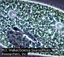
Paramecium
The paramecium is a single-celled organism that propels itself by minute, hairlike projections called cilia. Cilia also create currents that sweep food particles toward the paramecium’s gullet for ingestion.
M.I. Walker/Science Source/Photo Researchers, Inc.
Cell (biology), basic unit of life. Cells are the smallest structures capable of basic life processes, such as taking in nutrients, expelling waste, and reproducing. All living things are composed of cells. Some microscopic organisms, such as bacteria and protozoa, are unicellular, meaning they consist of a single cell. Plants, animals, and fungi are multicellular; that is, they are composed of a great many cells working in concert. But whether it makes up an entire bacterium or is just one of trillions in a human being, the cell is a marvel of design and efficiency. Cells carry out thousands of biochemical reactions each minute and reproduce new cells that perpetuate life.
Cells vary considerably in size. The smallest cell, a type of bacterium known as a mycoplasma, measures 0.0001 mm (0.000004 in) in diameter; 10,000 mycoplasmas in a row are only as wide as the diameter of a human hair. Among the largest cells are the nerve cells that run down a giraffe’s neck; these cells can exceed 3 m (9.7 ft) in length. Human cells also display a variety of sizes, from small red blood cells that measure 0.00076 mm (0.00003 in) to liver cells that may be ten times larger. About 10,000 average-sized human cells can fit on the head of a pin.
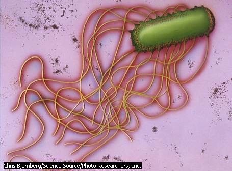
Along with their differences in size, cells present an array of shapes. Some, such as the bacterium Escherichia coli, resemble rods. The paramecium, a type of protozoan, is slipper shaped; and the amoeba, another protozoan, has an irregular form that changes shape as it moves around. Plant cells typically resemble boxes or cubes. In humans, the outermost layers of skin cells are flat, while muscle cells are long and thin. Some nerve cells, with their elongated, tentacle-like extensions, suggest an octopus.
In multicellular organisms, shape is typically tailored to the cell’s job. For example, flat skin cells pack tightly into a layer that protects the underlying tissues from invasion by bacteria. Long, thin muscle cells contract readily to move bones. The numerous extensions from a nerve cell enable it to connect to several other nerve cells in order to send and receive messages rapidly and efficiently.
By itself, each cell is a model of independence and self-containment. Like some miniature, walled city in perpetual rush hour, the cell constantly bustles with traffic, shuttling essential molecules from place to place to carry out the business of living. Despite their individuality, however, cells also display a remarkable ability to join, communicate, and coordinate with other cells. The human body, for example, consists of an estimated 20 to 30 trillion cells. Dozens of different kinds of cells are organized into specialized groups called tissues. Tendons and bones, for example, are composed of connective tissue, whereas skin and mucous membranes are built from epithelial tissue. Different tissue types are assembled into organs, which are structures specialized to perform particular functions. Examples of organs include the heart, stomach, and brain. Organs, in turn, are organized into systems such as the circulatory, digestive, or nervous systems. All together, these assembled organ systems form the human body.
The components of cells are molecules, nonliving structures formed by the union of atoms. Small molecules serve as building blocks for larger molecules. Proteins, nucleic acids, carbohydrates, and lipids, which include fats and oils, are the four major molecules that underlie cell structure and also participate in cell functions. For example, a tightly organized arrangement of lipids, proteins, and protein-sugar compounds forms the plasma membrane, or outer boundary, of certain cells. The organelles, membrane-bound compartments in cells, are built largely from proteins. Biochemical reactions in cells are guided by enzymes, specialized proteins that speed up chemical reactions. The nucleic acid deoxyribonucleic acid (DNA) contains the hereditary information for cells, and another nucleic acid, ribonucleic acid(RNA), works with DNA to build the thousands of proteins the cell needs.
| II | CELL STRUCTURE |
Cells fall into one of two categories: prokaryotic or eukaryotic (see Prokaryote). In a prokaryotic cell, found only in bacteria and archaebacteria, all the components, including the DNA, mingle freely in the cell’s interior, a single compartment. Eukaryotic cells, which make up plants, animals, fungi, and all other life forms, contain numerous compartments, or organelles, within each cell. The DNA in eukaryotic cells is enclosed in a special organelle called the nucleus, which serves as the cell’s command center and information library. The term prokaryote comes from Greek words that mean “before nucleus” or “prenucleus,” while eukaryote means “true nucleus.”
| A | Prokaryotic Cells |
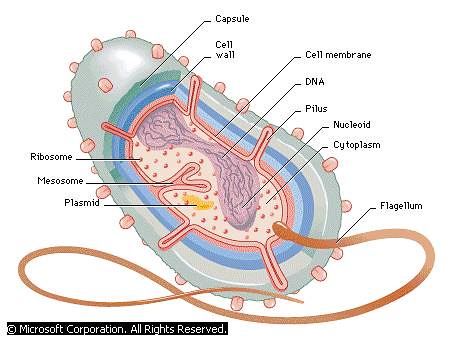
Anatomy of a Simple Bacterium
Bacteria cells typically are surrounded by a rigid, protective cell wall. The cell membrane, also called the plasma membrane, regulates passage of materials into and out of the cytoplasm, the semi-fluid that fills the cell. The DNA, located in the nucleoid region, contains the genetic information for the cell. Ribosomes carry out protein synthesis. Many baceteria contain a pilus (plural pili), a structure that extends out of the cell to transfer DNA to another bacterium. The flagellum, found in numerous species, is used for locomotion. Some bacteria contain a plasmid, a small chromososme with extra genes. Others have a capsule, a sticky substance external to the cell wall that protects bacteria from attack by white blood cells. Mesosomes were formerly thought to be structures with unknown functions, but now are know to be artifacts created when cells are prepared for viewing with electron microscopes.
© Microsoft Corporation. All Rights Reserved.
Prokaryotic cells are among the tiniest of all cells, ranging in size from 0.0001 to 0.003 mm (0.000004 to 0.0001 in) in diameter. About a hundred typical prokaryotic cells lined up in a row would match the thickness of a book page. These cells, which can be rodlike, spherical, or spiral in shape, are surrounded by a protective cell wall. Like most cells, prokaryotic cells live in a watery environment, whether it is soil moisture, a pond, or the fluid surrounding cells in the human body. Tiny pores in the cell wall enable water and the substances dissolved in it, such as oxygen, to flow into the cell; these pores also allow wastes to flow out.
Pushed up against the inner surface of the prokaryotic cell wall is a thin membrane called the plasma membrane. The plasma membrane, composed of two layers of flexible lipid molecules and interspersed with durable proteins, is both supple and strong. Unlike the cell wall, whose open pores allow the unregulated traffic of materials in and out of the cell, the plasma membrane is selectively permeable, meaning it allows only certain substances to pass through. Thus, the plasma membrane actively separates the cell’s contents from its surrounding fluids.
While small molecules such as water, oxygen, and carbon dioxide diffuse freely across the plasma membrane, the passage of many larger molecules, including amino acids (the building blocks of proteins) and sugars, is carefully regulated. Specialized transport proteins accomplish this task. The transport proteins span the plasma membrane, forming an intricate system of pumps and channels through which traffic is conducted. Some substances swirling in the fluid around the cell can enter it only if they bind to and are escorted in by specific transport proteins. In this way, the cell fine-tunes its internal environment.
The plasma membrane encloses the cytoplasm, the semifluid that fills the cell. Composed of about 65 percent water, the cytoplasm is packed with up to a billion molecules per cell, a rich storehouse that includes enzymes and dissolved nutrients, such as sugars and amino acids. The water provides a favorable environment for the thousands of biochemical reactions that take place in the cell.
Within the cytoplasm of all prokaryotes is deoxyribonucleic acid (DNA), a complex molecule in the form of a double helix, a shape similar to a spiral staircase. The DNA is about 1,000 times the length of the cell, and to fit inside, it repeatedly twists and folds to form a compact structure called a chromosome. The chromosome in prokaryotes is circular, and is located in a region of the cell called the nucleoid. Often, smaller chromosomes called plasmids are located in the cytoplasm. The DNA is divided into units called genes, just like a long train is divided into separate cars. Depending on the species, the DNA contains several hundred or even thousands of genes. Typically, one gene contains coded instructions for building all or part of a single protein. Enzymes, which are specialized proteins, determine virtually all the biochemical reactions that support and sustain the cell.
Also immersed in the cytoplasm are the only organelles in prokaryotic cells—tiny bead-like structures called ribosomes. These are the cell’s protein factories. Following the instructions encoded in the DNA, ribosomes churn out proteins by the hundreds every minute, providing needed enzymes, the replacements for worn-out transport proteins, or other proteins required by the cell.
While relatively simple in construction, prokaryotic cells display extremely complex activity. They have a greater range of biochemical reactions than those found in their larger relatives, the eukaryotic cells. The extraordinary biochemical diversity of prokaryotic cells is manifested in the wide-ranging lifestyles of the archaebacteria and the bacteria, whose habitats include polar ice, deserts, and hydrothermal vents—deep regions of the ocean under great pressure where hot water geysers erupt from cracks in the ocean floor.
| B | Eukaryotic Animal Cells |
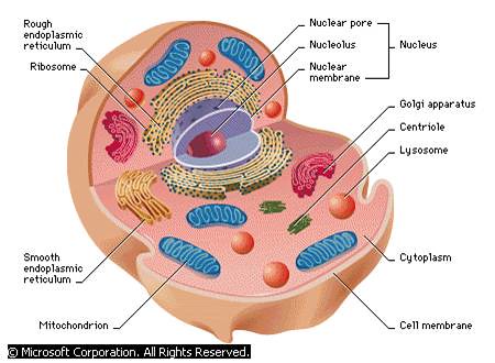
Animal Cell
An animal cell typically contains several types of membrane-bound organs, or organelles. The nucleus directs activities of the cell and carries genetic information from generation to generation. The mitochondria generate energy for the cell. Proteins are manufactured by ribosomes, which are bound to the rough endoplasmic reticulum or float free in the cytoplasm. The Golgi apparatus modifies, packages, and distributes proteins while lysosomes store enzymes for digesting food. The entire cell is wrapped in a lipid membrane that selectively permits materials to pass in and out of the cytoplasm.
© Microsoft Corporation. All Rights Reserved.
Eukaryotic cells are typically about ten times larger than prokaryotic cells. In animal cells, the plasma membrane, rather than a cell wall, forms the cell’s outer boundary. With a design similar to the plasma membrane of prokaryotic cells, it separates the cell from its surroundings and regulates the traffic across the membrane.
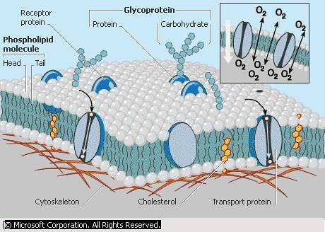
Plasma Membrane
The plasma membrane that surrounds eukaryotic cells is a dynamic structure composed of two layers of phospholipid molecules interspersed with cholesterol and proteins. Phospholipids are composed of a hydrophilic, or water-loving, head and two tails, which are hydrophobic, or water-hating. The two phospholipid layers face each other in the membrane, with the heads directed outward and the tails pointing inward. The water-attracting heads anchor the membrane to the cytoplasm, the watery fluid inside the cell, and also to the water surrounding the cell. The water-hating tails block large water-soluble molecules from passing through the membrane while permitting fat-soluble molecules, including medications such as tranquilizers and sleeping pills, to freely cross the membrane. Proteins embedded in the plasma membrane carry out a variety of functions, including transport of large water soluble molecules such as sugars and certain amino acids. Glycoproteins, proteins bonded to carbohydrates, serve in part to identify the cell as belonging to a unique organism, enabling the immune system to detect foreign cells, such as invading bacteria, which carry different glycoproteins. Cholesterol molecules in the plasma membrane act as stabilizers that limit the movement of the two slippery phospholipids layers, which slide back and forth in the membrane. Tiny gaps in the membrane enable small molecules such as oxygen (upper right) to diffuse readily into and out of the cell. Since cells constantly use up oxygen, decreasing its concentration within the cell, the higher concentration of oxygen outside the cell causes a net flow of oxygen into the cell. The steady stream of oxygen into the cell enables it to carry out aerobic respiration continually, a process that provides the cell with the energy needed to carry out its functions.
© Microsoft Corporation. All Rights Reserved.
The nucleus is the largest organelle in an animal cell. It contains numerous strands of DNA, the length of each strand being many times the diameter of the cell. Unlike the circular prokaryotic DNA, long sections of eukaryotic DNA pack into the nucleus by wrapping around proteins. As a cell begins to divide, each DNA strand folds over onto itself several times, forming a rod-shaped chromosome.
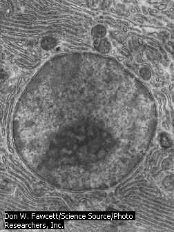
Nucleus of a Cell
The nucleus, present in eukaryotic cells, is a discrete structure containing chromosomes, which hold the genetic information for the cell. Separated from the cytoplasm of the cell by a double-layered membrane called the nuclear envelope, the nucleus contains a cellular material called nucleoplasm. Nuclear pores, present around the circumference of the nuclear membrane, allow the exchange of cellular materials between the nucleoplasm and the cytoplasm.
Don W. Fawcett/Science Source/Photo Researchers, Inc.
Attached to the nuclear membrane is an elongated membranous sac called the endoplasmic reticulum. This organelle tunnels through the cytoplasm, folding back and forth on itself to form a series of membranous stacks. Endoplasmic reticulum takes two forms: rough and smooth. Rough endoplasmic reticulum (RER) is so called because it appears bumpy under a microscope. The bumps are actually thousands of ribosomes attached to the membrane’s surface. The ribosomes in eukaryotic cells have the same function as those in prokaryotic cells—protein synthesis—but they differ slightly in structure. Eukaryote ribosomes bound to the endoplasmic reticulum help assemble proteins that typically are exported from the cell. The ribosomes work with other molecules to link amino acids to partially completed proteins. These incomplete proteins then travel to the inner chamber of the endoplasmic reticulum, where chemical modifications, such as the addition of a sugar, are carried out. Chemical modifications of lipids are also carried out in the endoplasmic reticulum.
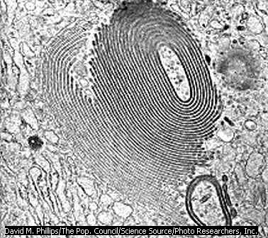
Golgi Apparatus
The Golgi apparatus, a minute cellular inclusion in the cytoplasm, is a series of smooth, stacked membranous sacs. The Golgi apparatus modifies proteins after they are produced by the ribosomes.
David M. Phillips/The Pop. Council/Science Source/Photo Researchers, Inc.
The second form of endoplasmic reticulum, the smooth endoplasmic reticulum (SER), lacks ribosomes and has an even surface. Within the winding channels of the smooth endoplasmic reticulum are the enzymes needed for the construction of molecules such as carbohydrates and lipids. The smooth endoplasmic reticulum is prominent in liver cells, where it also serves to detoxify substances such as alcohol, drugs, and other poisons.
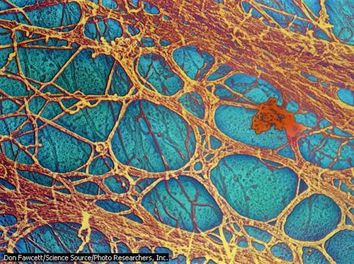
Cytoskeleton
The cytoskeleton, a network of protein fibers, crisscrosses the cytoplasm of eukaryotic cells, providing shape and mechanical support. The cytoskeleton also functions as a monorail to transport substances around the cell. A cell such as an amoeba changes shape by dismantling parts of the cytoskeleton and reassembling them in other locations.
Don Fawcett/Science Source/Photo Researchers, Inc.
Lysosomes are small, often spherical organelles that function as the cell’s recycling center and garbage disposal. Powerful digestive enzymes concentrated in the lysosome break down worn-out organelles and ship their building blocks to the cytoplasm where they are used to construct new organelles. Lysosomes also dismantle and recycle proteins, lipids, and other molecules.
The mitochondria are the powerhouses of the cell. Within these long, slender organelles, which can appear oval or bean shaped under the electron microscope, enzymes convert the sugar glucose and other nutrients into adenosine triphosphate (ATP). This molecule, in turn, serves as an energy battery for countless cellular processes, including the shuttling of substances across the plasma membrane, the building and transport of proteins and lipids, the recycling of molecules and organelles, and the dividing of cells. Muscle and liver cells are particularly active and require dozens and sometimes up to a hundred mitochondria per cell to meet their energy needs. Mitochondria are unusual in that they contain their own DNA in the form of a prokaryote-like circular chromosome; have their own ribosomes, which resemble prokaryotic ribosomes; and divide independently of the cell.
Unlike the tiny prokaryotic cell, the relatively large eukaryotic cell requires structural support. The cytoskeleton, a dynamic network of protein tubes, filaments, and fibers, crisscrosses the cytoplasm, anchoring the organelles in place and providing shape and structure to the cell. Many components of the cytoskeleton are assembled and disassembled by the cell as needed. During cell division, for example, a special structure called a spindle is built to move chromosomes around. After cell division, the spindle, no longer needed, is dismantled. Some components of the cytoskeleton serve as microscopic tracks along which proteins and other molecules travel like miniature trains. Recent research suggests that the cytoskeleton also may be a mechanical communication structure that converses with the nucleus to help organize events in the cell.
| C | Eukaryotic Plant Cells |
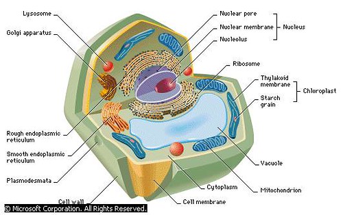
Plant Cell
Plant cells contain a variety of membrane-bound structures called organelles. These include a nucleus that carries genetic material; mitochondria that generate energy; ribosomes that manufacture proteins; smooth endoplasmic reticulum that manufactures lipids used for making membranes and storing energy; and a thin lipid membrane that surrounds the cell. Plant cells also contain chloroplasts that capture energy from sunlight and a single fluid-filled vacuole that stores compounds and helps in plant growth. Plant cells are surrounded by a rigid cell wall that protects the cell and maintains its shape.
© Microsoft Corporation. All Rights Reserved.
Plant cells have all the components of animal cells and boast several added features, including chloroplasts, a central vacuole, and a cell wall. Chloroplasts convert light energy—typically from the Sun—into the sugar glucose, a form of chemical energy, in a process known as photosynthesis. Chloroplasts, like mitochondria, possess a circular chromosome and prokaryote-like ribosomes, which manufacture the proteins that the chloroplasts typically need.
The central vacuole of a mature plant cell typically takes up most of the room in the cell. The vacuole, a membranous bag, crowds the cytoplasm and organelles to the edges of the cell. The central vacuole stores water, salts, sugars, proteins, and other nutrients. In addition, it stores the blue, red, and purple pigments that give certain flowers their colors. The central vacuole also contains plant wastes that taste bitter to certain insects, thus discouraging the insects from feasting on the plant.
In plant cells, a sturdy cell wall surrounds and protects the plasma membrane. Its pores enable materials to pass freely into and out of the cell. The strength of the wall also enables a cell to absorb water into the central vacuole and swell without bursting. The resulting pressure in the cells provides plants with rigidity and support for stems, leaves, and flowers. Without sufficient water pressure, the cells collapse and the plant wilts.
| III | CELL FUNCTIONS |
To stay alive, cells must be able to carry out a variety of functions. Some cells must be able to move, and most cells must be able to divide. All cells must maintain the right concentration of chemicals in their cytoplasm, ingest food and use it for energy, recycle molecules, expel wastes, and construct proteins. Cells must also be able to respond to changes in their environment.
| A | Movement |
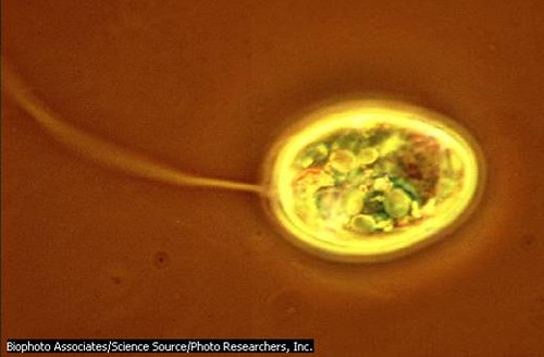
Euglena, Showing Whiplike Flagellum
The euglena is a single-celled alga with two or several flagella (depending on the species) located at one end for locomotion. Other algae, vertebrate sperm cells, and some protozoans and bacteria possess a single flagellum for movement.
Biophoto Associates/Science Source/Photo Researchers, Inc.
Movement in eukaryotes is also accomplished with cilia, short, hairlike proteins built by centrioles, which are barrel-shaped structures located in the cytoplasm that assemble and break down protein filaments. Typically, thousands of cilia extend through the plasma membrane and cover the surface of the cell, giving it a dense, hairy appearance. By beating its cilia as if they were oars, an organism such as the paramecium propels itself through its watery environment. In cells that do not move, cilia are used for other purposes. In the respiratory tract of humans, for example, millions of ciliated cells prevent inhaled dust, smog, and microorganisms from entering the lungs by sweeping them up on a current of mucus into the throat, where they are swallowed. Eukaryotic flagella and cilia are formed from basal bodies, small protein structures located just inside the plasma membrane. Basal bodies also help to anchor flagella and cilia.
Still other eukaryotic cells, such as amoebas and white blood cells, move by amoeboid motion, or crawling. They extrude their cytoplasm to form temporary pseudopodia, or false feet, which actually are placed in front of the cell, rather like extended arms. They then drag the trailing end of their cytoplasm up to the pseudopodia. A cell using amoeboid motion would lose a race to a euglena or paramecium. But while it is slow, amoeboid motion is strong enough to move cells against a current, enabling water-dwelling organisms to pursue and devour prey, for example, or white blood cells roaming the blood stream to stalk and engulf a bacterium or virus.
| B | Nutrition |
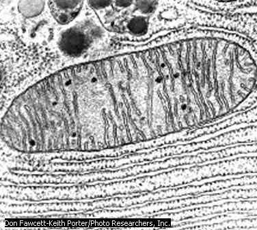
Mitochondria
Mitochondria, minute sausage-shaped structures found in the clear cytoplasm of the cell, are responsible for energy production. Mitochondria contain enzymes that help convert food material into adenosine triphosphate (ATP), which can be used directly by the cell as an energy source. Mitochondria tend to be concentrated near cellular structures that require large inputs of energy, such as the flagellum, which is responsible for movement in sperm cells and single-celled plants and animals.
Don Fawcett-Keith Porter/Photo Researchers, Inc.
Cells require energy for a variety of functions, including moving, building up and breaking down molecules, and transporting substances across the plasma membrane. Nutrients contains energy, but cells must convert the energy locked in nutrients to another form—specifically, the ATP molecule, the cell’s energy battery—before it is useful. In single-celled eukaryotic organisms, such as the paramecium, and in multicellular eukaryotic organisms, such as plants, animals, and fungi, mitochondria are responsible for this task. The interior of each mitochondrion consists of an inner membrane that is folded into a mazelike arrangement of separate compartments called cristae. Within the cristae, enzymes form an assembly line where the energy in glucose and other energy-rich nutrients is harnessed to build ATP; thousands of ATP molecules are constructed each second in a typical cell. In most eukaryotic cells, this process requires oxygen and is known as aerobic respiration.
Some prokaryotic organisms also carry out aerobic respiration. They lack mitochondria, however, and carry out aerobic respiration in the cytoplasm with the help of enzymes sequestered there. Many prokaryote species live in environments where there is little or no oxygen, environments such as mud, stagnant ponds, or within the intestines of animals. Some of these organisms produce ATP without oxygen in a process known as anaerobic respiration, where sulfur or other substances take the place of oxygen. Still other prokaryotes, and yeast, a single-celled eukaryote, build ATP without oxygen in a process known as fermentation.
Almost all organisms rely on the sugar glucose to produce ATP. Glucose is made by the process of photosynthesis, in which light energy is transformed to the chemical energy of glucose. Animals and fungi cannot carry out photosynthesis and depend on plants and other photosynthetic organisms for this task. In plants, as we have seen, photosynthesis takes place in organelles called chloroplasts. Chloroplasts contain numerous internal compartments called thylakoids where enzymes aid in the energy conversion process. A single leaf cell contains 40 to 50 chloroplasts. With sufficient sunlight, one large tree is capable of producing upwards of two tons of sugar in a single day. Photosynthesis in prokaryotic organisms—typically aquatic bacteria—is carried out with enzymes clustered in plasma membrane folds called chromatophores. Aquatic bacteria produce the food consumed by tiny organisms living in ponds, rivers, lakes, and seas.
| D | Protein Synthesis |
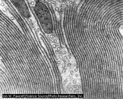
Ribosomes
On the surface of the rough endoplasmic reticulum are numerous small, dark structures called ribosomes. Ribosomes, which are also found floating free in the cytoplasm, are the sites of protein synthesis.
Don W. Fawcett/Science Source/Photo Researchers, Inc.
A typical cell must have on hand about 30,000 proteins at any one time. Many of these proteins are enzymes needed to construct the major molecules used by cells—carbohydrates, lipids, proteins, and nucleic acids—or to aid in the breakdown of such molecules after they have worn out. Other proteins are part of the cell’s structure—the plasma membrane and ribosomes, for example. In animals, proteins also function as hormones and antibodies, and they function like delivery trucks to transport other molecules around the body. Hemoglobin, for example, is a protein that transports oxygen in red blood cells. The cell’s demand for proteins never ceases.
Before a protein can be made, however, the molecular directions to build it must be extracted from one or more genes. In humans, for example, one gene holds the information for the protein insulin, the hormone that cells need to import glucose from the bloodstream, while at least two genes hold the information for collagen, the protein that imparts strength to skin, tendons, and ligaments. The process of building proteins begins when enzymes, in response to a signal from the cell, bind to the gene that carries the code for the required protein, or part of the protein. The enzymes transfer the code to a new molecule called messenger RNA, which carries the code from the nucleus to the cytoplasm. This enables the original genetic code to remain safe in the nucleus, with messenger RNA delivering small bits and pieces of information from the DNA to the cytoplasm as needed. Depending on the cell type, hundreds or even thousands of molecules of messenger RNA are produced each minute.
Once in the cytoplasm, the messenger RNA molecule links up with a ribosome. The ribosome moves along the messenger RNA like a monorail car along a track, stimulating another form of RNA—transfer RNA—to gather and link the necessary amino acids, pooled in the cytoplasm, to form the specific protein, or section of protein. The protein is modified as necessary by the endoplasmic reticulum and Golgi apparatus before embarking on its mission. Cells teem with activity as they forge the numerous, diverse proteins that are indispensable for life. For a more detailed discussion about protein synthesis, see Genetics: The Genetic Code.
| E | Cell Division |
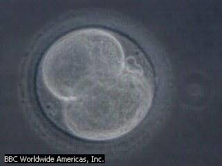
First Cell Divisions
As a fertilized egg goes through its first divisions, the daughter cells become progressively smaller. When there are a hundred or more cells, they form a hollow ball of cells, called a blastula, surrounding a fluid-filled cavity. Later divisions produce three layers of cells—endoderm (inner), mesoderm (middle), and ectoderm (outer)—from which the principal features of the animal will differentiate.
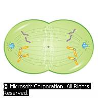
Mitosis
© Microsoft Corporation. All Rights Reserved.
Most cells divide at some time during their life cycle, and some divide dozens of times before they die. Organisms rely on cell division for reproduction, growth, and repair and replacement of damaged or worn out cells. Three types of cell division occur: binary fission, mitosis, and meiosis. Binary fission, the method used by prokaryotes, produces two identical cells from one cell. The more complex process of mitosis, which also produces two genetically identical cells from a single cell, is used by many unicellular eukaryotic organisms for reproduction. Multicellular organisms use mitosis for growth, cell repair, and cell replacement. In the human body, for example, an estimated 25 million mitotic cell divisions occur every second in order to replace cells that have completed their normal life cycles. Cells of the liver, intestine, and skin may be replaced every few days. Recent research indicates that even brain cells, once thought to be incapable of mitosis, undergo cell division in the part of the brain associated with memory.
The type of cell division required for sexual reproduction is meiosis. Sexually reproducing organisms include seaweeds, fungi, plants, and animals—including, of course, human beings. Meiosis differs from mitosis in that cell division begins with a cell that has a full complement of chromosomes and ends with gamete cells, such as sperm and eggs, that have only half the complement of chromosomes. When a sperm and egg unite during fertilization, the cell resulting from the union, called a zygote, contains the full number of chromosomes.
| IV | ORIGIN OF CELLS |

Fossilized Trilobites
Scientists study fossils to trace the evolution of life from simple cells to more complex organisms. Shown here are fossils of trilobites, primitive arthropods that once dominated the seas but became extinct about 250 million years ago.
James L. Amos/Photo Researchers, Inc.
The story of how cells evolved remains an open and actively investigated question in science (see Life). The combined expertise of physicists, geologists, chemists, and evolutionary biologists has been required to shed light on the evolution of cells from the nonliving matter of early Earth. The planet formed about 4.5 billion years ago, and for millions of years, violent volcanic eruptions blasted substances such as carbon dioxide, nitrogen, water, and other small molecules into the air. These small molecules, bombarded by ultraviolet radiation and lightning from intense storms, collided to form the stable chemical bonds of larger molecules, such as amino acids and nucleotides—the building blocks of proteins and nucleic acids. Experiments indicate that these larger molecules form spontaneously under laboratory conditions that simulate the probable early environment of Earth.
Scientists speculate that rain may have carried these molecules into lakes to create a primordial soup—a breeding ground for the assembly of proteins, the nucleic acid RNA, and lipids. Some scientists postulate that these more complex molecules formed in hydrothermal vents rather than in lakes. Other scientists propose that these key substances may have reached Earth on meteorites from outer space. Regardless of the origin or environment, however, scientists do agree that proteins, nucleic acids, and lipids provided the raw materials for the first cells. In the laboratory, scientists have observed lipid molecules joining to form spheres that resemble a cell’s plasma membrane. As a result of these observations, scientists postulate that millions of years of molecular collisions resulted in lipid spheres enclosing RNA, the simplest molecule capable of self-replication. These primitive aggregations would have been the ancestors of the first prokaryotic cells.
Fossil studies indicate that cyanobacteria, bacteria capable of photosynthesis, were among the earliest bacteria to evolve, an estimated 3.4 billion to 3.5 billion years ago. In the environment of the early Earth, there was no oxygen, and cyanobacteria probably used fermentation to produce ATP. Over the eons, cyanobacteria performed photosynthesis, which produces oxygen as a byproduct; the result was the gradual accumulation of oxygen in the atmosphere. The presence of oxygen set the stage for the evolution of bacteria that used oxygen in aerobic respiration, a more efficient ATP-producing process than fermentation. Some molecular studies of the evolution of genes in archaebacteria suggest that these organisms may have evolved in the hot waters of hydrothermal vents or hot springs slightly earlier than cyanobacteria, around 3.5 billion years ago. Like cyanobacteria, archaebacteria probably relied on fermentation to synthesize ATP.
Eukaryotic cells may have evolved from primitive prokaryotes about 2 billion years ago. One hypothesis suggests that some prokaryotic cells lost their cell walls, permitting the cell’s plasma membrane to expand and fold. These folds, ultimately, may have given rise to separate compartments within the cell—the forerunners of the nucleus and other organelles now found in eukaryotic cells. Another key hypothesis is known as endosymbiosis. Molecular studies of the bacteria-like DNA and ribosomes in mitochondria and chloroplasts indicate that mitochondrion and chloroplast ancestors were once free-living bacteria. Scientists propose that these free-living bacteria were engulfed and maintained by other prokaryotic cells for their ability to produce ATP efficiently and to provide a steady supply of glucose. Over generations, eukaryotic cells complete with mitochondria—the ancestors of animals—or with both mitochondria and chloroplasts—the ancestors of plants—evolved (see Evolution).
| V | THE DISCOVERY AND STUDY OF CELLS |
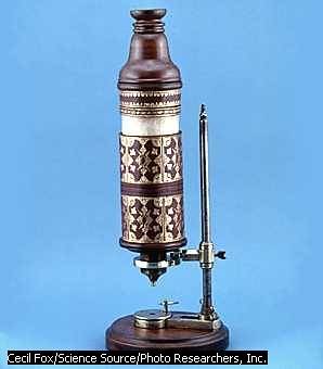
Hooke’s Microscope
English scientist Robert Hooke built this microscope in the 17th century and used it to conduct pioneering research. He discovered the cell structure of plants by observing a thin slice of cork under his microscope.
Cecil Fox/Science Source/Photo Researchers, Inc.
The first observations of cells were made in 1665 by English scientist Robert Hooke, who used a crude microscope of his own invention to examine a variety of objects, including a thin piece of cork. Noting the rows of tiny boxes that made up the dead wood’s tissue, Hooke coined the term cell because the boxes reminded him of the small cells occupied by monks in a monastery. While Hooke was the first to observe and describe cells, he did not comprehend their significance. At about the same time, the Dutch maker of microscopes Antoni van Leeuwenhoek pioneered the invention of one of the best microscopes of the time. Using his invention, Leeuwenhoek was the first to observe, draw, and describe a variety of living organisms, including bacteria gliding in saliva, one-celled organisms cavorting in pond water, and sperm swimming in semen. Two centuries passed, however, before scientists grasped the true importance of cells.
Modern ideas about cells appeared in the 1800s, when improved light microscopes enabled scientists to observe more details of cells. Working together, German botanist Matthias Jakob Schleiden and German zoologist Theodor Schwann recognized the fundamental similarities between plant and animal cells. In 1839 they proposed the revolutionary idea that all living things are made up of cells. Their theory gave rise to modern biology: a whole new way of seeing and investigating the natural world.

Antoni van Leeuwenhoek
Although lacking basic scientific training, Antoni van Leeuwenhoek is credited with inventing the precursor to the modern microscope. Leeuwenhoek was the first to document the structure of red blood corpuscles and the nature of the circulatory system. In addition to describing animalcules (protozoans and bacteria), Leeuwenhoek also accurately described the life cycles of many types of insects.
Culver Pictures
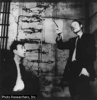
Francis Crick and James Watson
The deoxyribonucleic acid (DNA) molecule is the genetic blueprint for each cell and ultimately the blueprint that determines every characteristic of a living organism. In 1953 American biochemist James Watson, left, and British biophysicist Francis Crick, right, described the structure of the DNA molecule as a double helix, somewhat like a spiral staircase with many individual steps. Their work was aided by X-ray diffraction pictures of the DNA molecule taken by British biophysicist Maurice Wilkins and British physical chemist Rosalind Franklin. In 1962 Crick, Watson, and Wilkins received the Nobel Prize for their pioneering work on the structure of the DNA molecule.
Photo Researchers, Inc.
While some scientists were studying the functions of cells, others were examining details of their structure. They were aided by a crucial technological development in the 1940s: the invention of the electron microscope, which uses high-energy electrons instead of light waves to view specimens. New generations of electron microscopes have provided resolution, or the differentiation of separate objects, thousands of times more powerful than that available in light microscopes. This powerful resolution revealed organelles such as the endoplasmic reticulum, lysosomes, the Golgi apparatus, and the cytoskeleton. The scientific fields of cell structure and function continue to complement each other as scientists explore the enormous complexity of cells.

Scanning Electron Microscope
This scanning electron microscope (SEM) at the University of California, Berkeley is located to the left with the computer images of the specimen on the computer screens to the right. Although a SEM cannot resolve objects as small as a transmission electron microscope, the images produced by the SEM are more useful for seeing the three-dimensional aspect of the surface structure of small objects.
Lawrence Migdale/Photo Researchers, Inc.
Another busy area in cell biology concerns programmed cell death, or apoptosis. Millions of times per second in the human body, cells commit suicide as an essential part of the normal cycle of cellular replacement. This also seems to be a check against disease: When mutations build up within a cell, the cell will usually self-destruct. If this fails to occur, the cell may divide and give rise to mutated daughter cells, which continue to divide and spread, gradually forming a growth called a tumor. This unregulated growth by rogue cells can be benign, or harmless, or cancerous, which may threaten healthy tissue. The study of apoptosis is one avenue that scientists explore in an effort to understand how cells become cancerous.
Scientists are also discovering exciting aspects of the physical forces within cells. Cells employ a form of architecture called tensegrity, which enables them to withstand battering by a variety of mechanical stresses, such as the pressure of blood flowing around cells or the movement of organelles within the cell. Tensegrity stabilizes cells by evenly distributing mechanical stresses to the cytoskeleton and other cell components. Tensegrity also may explain how a change in the cytoskeleton, where certain enzymes are anchored, initiates biochemical reactions within the cell, and can even influence the action of genes. The mechanical rules of tensegrity may also account for the assembly of molecules into the first cells. Such new insights—made some 300 years after the tiny universe of cells was first glimpsed—show that cells continue to yield fascinating new worlds of discovery.
Contributed By:
Christopher King


0 komentar:
Posting Komentar